Tag: arm wrestling

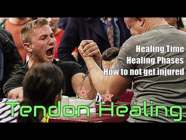
VIDEO: Tendon Healing (How to use tendon healing times in Arm Wrestling)

Tendon Healing

How is Tendinopathy Treated?

In most cases, you can start treating a tendon injury at home. To get the best results, start these steps right away:

  • Rest the painful area, and avoid any activity that makes the pain worse.
  • Apply ice or cold packs for 20 minutes at a time, as often as 2 times an hour, for the first 72 hours. Keep using ice as long as it helps.
  • Do gentle range-of-motion exercises and stretching to prevent stiffness.
  • Have your biomechanics assessed by a sports physiotherapist.
  • Undertake an Eccentric Strengthen Program. This is vital!

Tendon healing process

Tendon healing can be largely divided into 3 overlapping phases, inflammatory repairing and remodelling phases:

The initial inflammatory phase, which lasts about 24 hours, erythrocytes, platelets and inflammatory cells (eg: neutrophils, monocytes and macrophages) migrate to the wound site and clean the site of necrotic materials by phagocytosis.  In the meantime, these cells release phaso active and chemo tactic factors which recruit tendon fibroblast to begin collegan synthesis and deposition.

A few days after the injury, the repairing phase begins.  In this phase, which lasts a few weeks, tendon fibroblast synthesise abundant collegan and other extra cellular matrix components such as proteoglycans and deposit them at the wound site.

After about 6 weeks, the remodelling phase starts.  This phase is characterised by decreased cellularity and decreased collagen and glycosaminoglycan synthesis.  During this period, the repair tissue changes to fibrous tissue, this again changes to scar like tendon tissue after 10 weeks.  During the later remodelling phase covalent bonding between the collagen fibres increases resulting in repaired tissue with highest stiffness and tense our strength.  Also, both the metabolism of tenocytes and tendon vascularity decline.

During tissue healing growth factors play an important role in this process.

1:   Platelet Derived Growth Factor (PDGF) is produced shortly after tendon injury and stimulates the production of other growth factors.

2:  TGF-beta is active during the inflammatory and repair phases of tendon healing.  TGF-beta plays a major role in the repair of injured tendons.  TGF-beta 1 aids an extra cellular matrix deposition; however, it’s over expression results in tissue fibrosis.  TGF-beta 2 functions similarly to TGF-beta 1.  However, TGF-beta 3 has been shown to improve tissue scarring.  Peak levels of TGF-beta receptory expression occur at day 14 post injury and decrease until day 56 post injury.

  1. Vascular Endothelial Growth Factor (VEGF) stimulates endothelial cell proliferation, enhances angiogenesis and increases capillary permeability.  VEGF RNA expression is detected at the repair site 7 days post injury with peak levels at 10 days post injury.
  2. Nitric Oxide Synthase (NOS) isoforms are expressed with differential expression patterns during the 3 phases of tendon healing.

It should be noted that, except for degenerative tendons (tendonosis), injured tendons tend to heal. However, the healing tendon does not reach the biomechanical properties of the tendon prior to surgery.

VIDEOS: How to not to break your arm while armwrestling

Subscribe to channel for more armwrestling videos


This is most common place where injuries happen and most common mistake while armwrestling. Tips from pros on how to not get your arm broken in armwrestling.

Down is compilation video on arm breaks in armwrestling. All these arm breaks could have been avoided. Knowing armwrestling technique would decrease your chances of getting injured or your arm broken.

Some things that are common in all theses arm break videos:

– Pushing not Pulling

– Facing away from arm

– Moving Shoulder in front of arm


https://www.youtube.com/watch?v=VRYQf2TNm-w

Don`t take ibuprofen to stop inflammation for Arm Wrestling. This is killing your gains.

Many physicians consider NSAIDs to be the medication of choice for managing musculoskeletal pain and injury. However, studies have questioned their value in the healing process of bone, muscle, tendon, and ligament injuries and their use carries the risk of potentially serious adverse effects. Animal and human studies have linked NSAID use to poor fracture healing. There appears to be little role for NSAIDs in tendinopathy outside of initial symptomatic pain relief. Animal studies provide conflicting evidence of efficacy in ligament injury, but human trials suggest that short courses may be of benefit in acute injury. Experimental animal models mostly demonstrate no effect on muscle healing or a reduction in muscle strength. Alternatives for analgesia in musculoskeletal injuries include acetaminophen, opiate-containing medication, and topical preparations. (J Musculoskel Med. 2011;28:207-212)

Tendon injury
The treatment goal for tendinopathy, as for fractures, is decreased pain and return to normal function. The term “tendinopathy” has been associated with both chronic tendon degeneration (tendinosis) and acute injury (tendinitis). The majority of tendon disorders are deemed to be chronic, degenerative changes (tendinosis rather than tendinitis) and acute tendon injury resulting from overloading of tissue that already has undergone degenerative changes.

Studies have demonstrated that prostaglandins and leukotrienes are produced during the acute phase of a tendon injury and may be involved in the subsequent degenerative changes over the long term. In the first few days after acute tendon injury, there is an initial inflammatory phase with angiogenesis, increased vascular permeability, and entry of inflammatory cells into the injury site. Prostaglandins are thought to be involved in these processes.

How Not to Treat Acute Inflammation

  1. If you’re experiencing acute inflammation – the type that occurs after an injury, after surgery, or after a muscle-fiber damaging workout – avoid taking any NSAIDS. While they’ll help quell the pain, they impede the healing process and in the case of muscles, they may well impede further growth. Granted, the occasional use of NSAIDS won’t likely pose much of a problem, but if you take them regularly, the odds are high that you’re impeding muscle growth.
  2. Reconsider icing sore muscles. The initial thinking was that it was safe to ice a muscle because there was no need for immune cells to get all angried up when the injury was internal and there was no possibility of infection. The trouble is, that’s short sighted. Granted, icing will help with pain, but since we now know that inflammation is necessary for muscles to grow bigger and stronger, icing is likely counterproductive.

Instead, Treat Acute Inflammation Like This…

  1. If you’re hurting from acute inflammation caused by an injury or you’re debilitated from a brutal workout and you simply can’t handle it, consider using acetaminophen. While it does exhibit some anti-inflammatory effects, it’s more of an analgesic.
  2. If pain persists after a couple of days, take any NSAID you feel is necessary, or, preferably, take appropriate doses of Curcumin. Curcumin is a potent anti-inflammatory and it blocks a host of inflammatory compounds by a certain reasonable percentage, as opposed to blocking one particular inflammatory compound by 100 percent. As an example of the latter, the prescription painkiller Vioxx shuts down COX-2 production completely and got lots of bad press for causing all those pesky heart attacks.

VIDEO: Armwrestling Style and Arm Length (Long Arms and Short Arms)

Have you ever wondered if you practice the correct Armwrestling style for your Arm Length?
It’s often that we decide how to pull in the middle of the competition, and often the choice is wrong!

Prepare and preplan of how to use your advantages.

WATCH CLOSELY!

Consider subscribing if you want to be up to date with the newest videos containing exclusive value: SUBSCRIBE

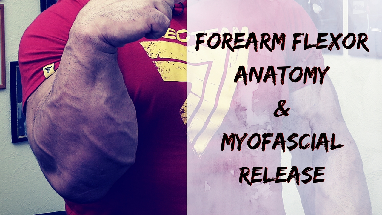
VIDEO: Forearm Anatomy and Flexor muscle Myofascial Release and Massage Techniques for Armwrestling

These muscles are largely involved with pronation. The superficial muscles have their origin on the common flexor tendon. The ulnar nerve and artery are also contained within this compartment.The flexor digitorum superficialis lies in between the other four muscles of the superficial group and the three muscles of the deep group. This is why it is also classified as the intermediate group.
Pain in different place of your arm can be caused by a lot of factors. But if pain is located on your muscle there is a big chance it`s a tight spot or trigger point. Armwrestling involves a lot of same movements from gripping (wrist and finger flexion). Any kind of moment that has been overdone can cause muscles to get tight. In this video we are showing some ideas about how to do self massage on your forearm flexor muscles using specific equipment and using things you can find in almost any gym. If your forearms get tight try these and leave a comment did it help.

For more Armwrestling videos click this button:

The muscles of the forearm can be divided into two groups: anterior (flexors) and posterior (extensors).Both the flexors and extensors are further divided into superficial and deep layers.The forearm muscles that control the movement of the hands are known as extrinsic hand muscles. These muscles originate outside the hand and insert on structures within it.

Shown here, the extrinsic hand muscles are the flexor carpi radialis, palmaris longis, flexor carpi ulnaris, and flexor digitorum superficialis.These muscles move the wrist, hand, fingers and thumb.The pronator teres inserts on the radius and pronates the forearm and hand.

The superficial muscles in the anterior compartment are the flexor carpi ulnaris, palmaris longus, flexor carpi radialis and pronator teres.  They all originate from a common tendon, which arises from the medial epicondyle of the humerus.

Flexor Carpi Ulnaris

  • Attachments:  Originates from the medial epicondyle with the other superficial flexors. It also has a long origin from the ulna. It passes into the wrist, and attaches to the pisiform carpal bone.
  • Actions: Flexion and adduction at the wrist.
  • Innervation: Ulnar nerve.

Palmaris Longus

This muscle is absent in about 15% of the population.

Dissection Tip: Just distal to the wrist, if you reflect back the palmaris longus, you will find the median nerve immediately underneath it

  • Attachments:  Originates from the medial epicondyle, attaches to the flexor retinaculum of the wrist.
  • Actions: Flexion at the wrist.
  • Innervation: Median nerve.

Flexor Carpi Radialis

  • Attachments: Originates from the medial epicondyle, attaches to the base of metacarpals II and III.
  • Actions: Flexion and abduction at the wrist.
  • Innervation: Median nerve.

Pronator Teres

The lateral border of the pronator teres forms the medial border of the cubital fossa, an anatomical triangle located over the elbow.

  • Attachments: It has two origins, one from the medial epicondyle, and the other from the coronoid process of the ulna. It attaches laterally to the mid-shaft of the radius.
  • Actions: Pronation of the forearm.
  • InnervationMedian nerve.

The flexor digitorum superficialis is the only muscle of the intermediate compartment. It can sometimes be classed as a superficial muscle, but in most cadavers it lies between the deep and superficial muscle layers.

The muscle is a good anatomical landmark in the forearm – the median nerve and ulnar artery pass between its two heads, and then travel posteriorly.

  • Attachments: It has two heads – one originates from the medial epicondyle of the humerus, the other from the radius. The muscle splits into four tendons at the wrist, which travel through the carpal tunnel, and attaches to the middle phalanges of the four fingers.
  • Actions: Flexes the metacarpophalangeal joints and proximal interphalangeal joints at the 4 fingers, and flexes at the wrist.
  • Innervation: Median nerve.

There are three muscles in the deep anterior forearm; flexor digitorum profundus, flexor pollicis longus, and pronator quadratus.

Flexor Digitorum Profundus

  • Attachments: Originates from the ulna and associated interosseous membrane. At the wrist, it splits into four tendons, that pass through the carpal tunnel and attach to the distal phalanges of the four fingers.
  • Actions: It is the only muscle that can flex the distal interphalangeal joints of the fingers. It also flexes at metacarpophalangeal joints and at the wrist.
  • Innervation: The medial half (acts on the little and ring fingers) is innervated by the ulnar nerve. The lateral half (acts on the middle and index fingers) is innervated by the anterior interosseous branch of the median nerve.

Flexor Pollicis Longus

This muscle lies laterally to the FDP.

  • Attachments: Originates from the anterior surface of the radius, and surrounding interosseous membrane. Attaches to the base of the distal phalanx of the thumb.
  • Actions:  Flexes the interphalangeal joint and metacarpophalangeal joint of the thumb.
  • Innervation: Median nerve (anterior interosseous branch).

Pronator Quadratus

A square shaped muscle, found deep to the tendons of the FDP and FPL.

  • Attachments: Originates from the anterior surface of the ulna, and attaches to the anterior surface of the radius.
  • Actions: Pronates the forearm.
  • Innervation: Median nerve (anterior interosseous branch).

Myofascial release (MFR, self-myofascial release) is an alternative medicine therapy that claims to treat skeletal muscle immobility and pain by relaxing contracted muscles, improving blood and lymphatic circulation, and stimulating the stretch reflex in muscles.

Fascia is a thin, tough, elastic type of connective tissue that wraps most structures within the human body, including muscle. Fascia supports and protects these structures. Osteopathic theory proposes that this soft tissue can become restricted due to psychogenic disease, overuse, trauma, infectious agents, or inactivity, often resulting in pain, muscle tension, and corresponding diminished blood flow.

Myofascial release focuses on reducing pain by easing the tension and tightness in the trigger points. It’s not always easy to understand what trigger point is responsible for the pain. Localizing pain to a specific trigger point is very difficult. For that reason, myofascial release is often used over a broad area of muscle and tissue rather than at single points.

All things I`m using in video you can find by clicking on picture.

FOAM ROLLER

Nano Triggerpoint Roller

Myofascial Release Ball

For more Armwrestling videos click this button:

https://youtu.be/JtfHw8NyEGw


Raimonds Liepiņš – Coach RayX

INSTAGRAM: https://www.instagram.com/coach_rayx/
FACEBOOK: https://www.facebook.com/coach.rayx
TWITTER: https://twitter.com/RaimondsLiepins