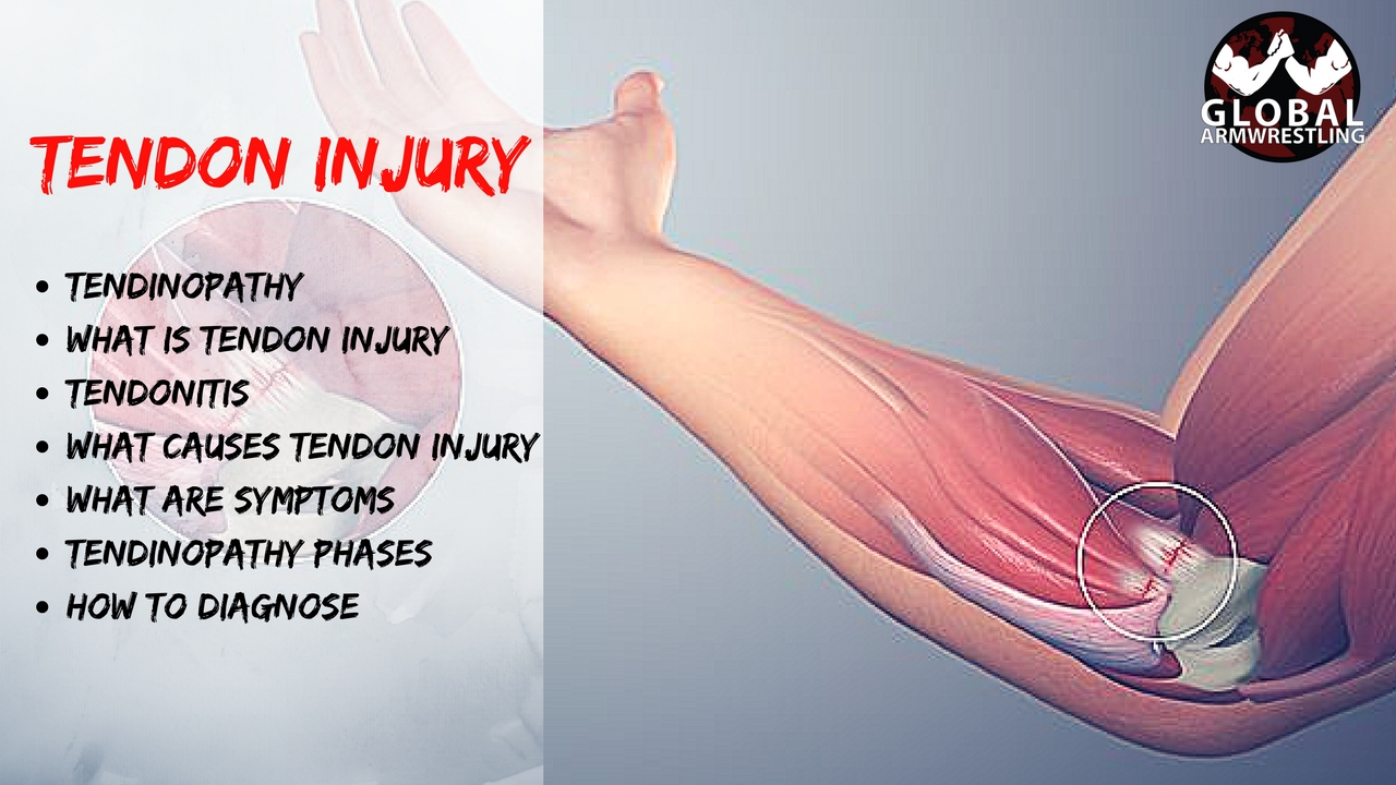
Category: ANATOMY

VIDEO: Tendon Healing (How to use tendon healing times in Arm Wrestling)

Tendon Healing

How is Tendinopathy Treated?

In most cases, you can start treating a tendon injury at home. To get the best results, start these steps right away:

  • Rest the painful area, and avoid any activity that makes the pain worse.
  • Apply ice or cold packs for 20 minutes at a time, as often as 2 times an hour, for the first 72 hours. Keep using ice as long as it helps.
  • Do gentle range-of-motion exercises and stretching to prevent stiffness.
  • Have your biomechanics assessed by a sports physiotherapist.
  • Undertake an Eccentric Strengthen Program. This is vital!

Tendon healing process

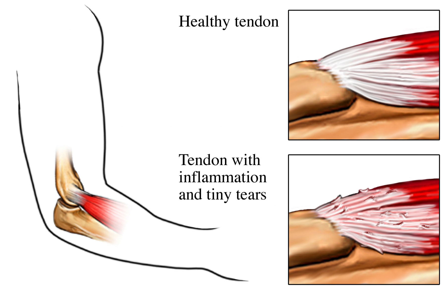
Tendon healing can be largely divided into 3 overlapping phases, inflammatory repairing and remodelling phases:

The initial inflammatory phase, which lasts about 24 hours, erythrocytes, platelets and inflammatory cells (eg: neutrophils, monocytes and macrophages) migrate to the wound site and clean the site of necrotic materials by phagocytosis.  In the meantime, these cells release phaso active and chemo tactic factors which recruit tendon fibroblast to begin collegan synthesis and deposition.

A few days after the injury, the repairing phase begins.  In this phase, which lasts a few weeks, tendon fibroblast synthesise abundant collegan and other extra cellular matrix components such as proteoglycans and deposit them at the wound site.

After about 6 weeks, the remodelling phase starts.  This phase is characterised by decreased cellularity and decreased collagen and glycosaminoglycan synthesis.  During this period, the repair tissue changes to fibrous tissue, this again changes to scar like tendon tissue after 10 weeks.  During the later remodelling phase covalent bonding between the collagen fibres increases resulting in repaired tissue with highest stiffness and tense our strength.  Also, both the metabolism of tenocytes and tendon vascularity decline.

During tissue healing growth factors play an important role in this process.

1:   Platelet Derived Growth Factor (PDGF) is produced shortly after tendon injury and stimulates the production of other growth factors.

2:  TGF-beta is active during the inflammatory and repair phases of tendon healing.  TGF-beta plays a major role in the repair of injured tendons.  TGF-beta 1 aids an extra cellular matrix deposition; however, it’s over expression results in tissue fibrosis.  TGF-beta 2 functions similarly to TGF-beta 1.  However, TGF-beta 3 has been shown to improve tissue scarring.  Peak levels of TGF-beta receptory expression occur at day 14 post injury and decrease until day 56 post injury.

  1. Vascular Endothelial Growth Factor (VEGF) stimulates endothelial cell proliferation, enhances angiogenesis and increases capillary permeability.  VEGF RNA expression is detected at the repair site 7 days post injury with peak levels at 10 days post injury.
  2. Nitric Oxide Synthase (NOS) isoforms are expressed with differential expression patterns during the 3 phases of tendon healing.

It should be noted that, except for degenerative tendons (tendonosis), injured tendons tend to heal. However, the healing tendon does not reach the biomechanical properties of the tendon prior to surgery.

VIDEOS: How to not to break your arm while armwrestling

Subscribe to channel for more armwrestling videos


This is most common place where injuries happen and most common mistake while armwrestling. Tips from pros on how to not get your arm broken in armwrestling.

Down is compilation video on arm breaks in armwrestling. All these arm breaks could have been avoided. Knowing armwrestling technique would decrease your chances of getting injured or your arm broken.

Some things that are common in all theses arm break videos:

– Pushing not Pulling

– Facing away from arm

– Moving Shoulder in front of arm


https://www.youtube.com/watch?v=VRYQf2TNm-w

VIDEO: Tendinopathy (Tendon Injury)

Tendinopathy (tendon injuries) can develop in any tendon of the body.

Typically, tendon injuries occur in three areas:

  • musculotendinous junction (where the tendon joins the muscle)
  • mid-tendon (non-insertional tendinopathy)
  • tendon insertion (eg into bone)

Non-insertional tendinopathies tends to be caused by a cumulative microtrauma from repetitive overloading eg overtraining.


What is a Tendon Injury?

Tendons are the tough fibres that connect muscle to bone. Most tendon injuries occur near joints, such as the shoulder, elbow, knee, and ankle. A tendon injury may seem to happen suddenly, but usually, it is the result of repetitive tendon overloading. Health professionals may use different terms to describe a tendon injury. You may hear:

Tendinitis (or Tendonitis): This actually means “inflammation of the tendon,” but inflammation is actually normal tendon healing response which can cause some tendon pain. This is known as the reactive phase and is a good tendon healing response.

The problem really occurs when you healing rate is less than your injury rate – known as tendon dysrepair – which is when tendinopathies can quickly deteriorate into the degenerative (cell death) phase. This is characterized by collagen degeneration in the tendon due to repetitive overloading. These tendinopathies therefore do not respond well to anti-inflammatory treatments and are best treated with functional rehabilitation. The best results occur with early diagnosis and intervention.

What Causes a Tendon Injury?

Most tendon injuries are the result of gradual wear and tear to the tendon from overuse or ageing. Anyone can have a tendon injury, but people who make the same motions over and over in their jobs, sports, or daily activities are more likely to damage a tendon.

Your tendons are designed to withstand high, repetitive loading, however, on occasions, when the load being applied to the tendon is too great for the tendon to withstand, the tendon begins to become stressed.

When tendons become stressed, they sustain small micro tears, which encourage inflammatory chemicals and swelling, which can quickly heal if managed appropriately.

However, if the load is continually applied to the tendon, these lesions occurring in the tendon can exceed the rate of repair. The damage will progressively become worse, causing pain and dysfunction. The result is a tendinopathy or tendinosis.

Researchers current opinion implicates the cumulative microtrauma associated with high tensile and compressive forces generated during sport or an activity causes a tendinopathy.

For example, in explosive jumping movements, forces delivered to the patellar tendon can be eight times your body weight. Cumulative micro trauma appears to exceed the tendon’s capacity to heal and remodel.


What are the Symptoms of Tendinopathy?

Tendinopathy usually causes pain, stiffness, and loss of strength in the affected area.

  • The pain may get worse when you use the tendon.
  • You may have more pain and stiffness during the night or when you get up in the morning.
  • The area may be tender, red, warm, or swollen if there is inflammation.
  • You may notice a crunchy sound or feeling when you use the tendon.

The symptoms of a tendon injury can be a lot like those caused by bursitis.

Tendinopathy Phases

The inability of your tendon to adapt to the load quickly enough causes tendon to progress through four phases of tendon injury. While it is healthy for normal tissue adaptation during phase one, further progression can lead to tendon cell death and subsequent tendon rupture.

1. Reactive Tendinopathy

  • Normal tissue adaptation phase
  • Prognosis: Excellent. Normal Recovery!

2. Tendon Dysrepair

  • Injury rate > Repair rate
  • Prognosis: Good. Tissue is attempting to heal.
  • It is vital that you prevent deterioration and progression to permanent cell death (phase 3).

3. Degenerative Tendinopathy

  • Cell death occurs
  • Poor Prognosis – Tendon cells are giving up!

4. Tendon Tear or Rupture

  • Catastrophic tissue breakdown
  • Loss of function.
  • Prognosis: very poor.
  • Surgery is often the only option.

It is very important to have your tendinopathy professionally assessed to identify it’s injury phase. Identifying your tendinopathy phase is also vital to direct your most effective treatment, since certain modalities or exercises should only be applied or undertaken in specific tendon healing phases.

How is a Tendon Injury Diagnosed?

To diagnose a tendon injury, your physiotherapist will ask questions about your past health, your symptoms and exercise regime. They’ll then do a physical examination to confirm the diagnosis. If your symptoms are severe or you do not improve with early treatment, specific diagnostic tests may be requested, such as an ultrasound scan or MRI.

For more Armwrestling videos click this button:

VIDEO: Forearm Anatomy and Flexor muscle Myofascial Release and Massage Techniques for Armwrestling

These muscles are largely involved with pronation. The superficial muscles have their origin on the common flexor tendon. The ulnar nerve and artery are also contained within this compartment.The flexor digitorum superficialis lies in between the other four muscles of the superficial group and the three muscles of the deep group. This is why it is also classified as the intermediate group.
Pain in different place of your arm can be caused by a lot of factors. But if pain is located on your muscle there is a big chance it`s a tight spot or trigger point. Armwrestling involves a lot of same movements from gripping (wrist and finger flexion). Any kind of moment that has been overdone can cause muscles to get tight. In this video we are showing some ideas about how to do self massage on your forearm flexor muscles using specific equipment and using things you can find in almost any gym. If your forearms get tight try these and leave a comment did it help.

For more Armwrestling videos click this button:

The muscles of the forearm can be divided into two groups: anterior (flexors) and posterior (extensors).Both the flexors and extensors are further divided into superficial and deep layers.The forearm muscles that control the movement of the hands are known as extrinsic hand muscles. These muscles originate outside the hand and insert on structures within it.

Shown here, the extrinsic hand muscles are the flexor carpi radialis, palmaris longis, flexor carpi ulnaris, and flexor digitorum superficialis.These muscles move the wrist, hand, fingers and thumb.The pronator teres inserts on the radius and pronates the forearm and hand.

The superficial muscles in the anterior compartment are the flexor carpi ulnaris, palmaris longus, flexor carpi radialis and pronator teres.  They all originate from a common tendon, which arises from the medial epicondyle of the humerus.

Flexor Carpi Ulnaris

  • Attachments:  Originates from the medial epicondyle with the other superficial flexors. It also has a long origin from the ulna. It passes into the wrist, and attaches to the pisiform carpal bone.
  • Actions: Flexion and adduction at the wrist.
  • Innervation: Ulnar nerve.

Palmaris Longus

This muscle is absent in about 15% of the population.

Dissection Tip: Just distal to the wrist, if you reflect back the palmaris longus, you will find the median nerve immediately underneath it

  • Attachments:  Originates from the medial epicondyle, attaches to the flexor retinaculum of the wrist.
  • Actions: Flexion at the wrist.
  • Innervation: Median nerve.

Flexor Carpi Radialis

  • Attachments: Originates from the medial epicondyle, attaches to the base of metacarpals II and III.
  • Actions: Flexion and abduction at the wrist.
  • Innervation: Median nerve.

Pronator Teres

The lateral border of the pronator teres forms the medial border of the cubital fossa, an anatomical triangle located over the elbow.

  • Attachments: It has two origins, one from the medial epicondyle, and the other from the coronoid process of the ulna. It attaches laterally to the mid-shaft of the radius.
  • Actions: Pronation of the forearm.
  • InnervationMedian nerve.

The flexor digitorum superficialis is the only muscle of the intermediate compartment. It can sometimes be classed as a superficial muscle, but in most cadavers it lies between the deep and superficial muscle layers.

The muscle is a good anatomical landmark in the forearm – the median nerve and ulnar artery pass between its two heads, and then travel posteriorly.

  • Attachments: It has two heads – one originates from the medial epicondyle of the humerus, the other from the radius. The muscle splits into four tendons at the wrist, which travel through the carpal tunnel, and attaches to the middle phalanges of the four fingers.
  • Actions: Flexes the metacarpophalangeal joints and proximal interphalangeal joints at the 4 fingers, and flexes at the wrist.
  • Innervation: Median nerve.

There are three muscles in the deep anterior forearm; flexor digitorum profundus, flexor pollicis longus, and pronator quadratus.

Flexor Digitorum Profundus

  • Attachments: Originates from the ulna and associated interosseous membrane. At the wrist, it splits into four tendons, that pass through the carpal tunnel and attach to the distal phalanges of the four fingers.
  • Actions: It is the only muscle that can flex the distal interphalangeal joints of the fingers. It also flexes at metacarpophalangeal joints and at the wrist.
  • Innervation: The medial half (acts on the little and ring fingers) is innervated by the ulnar nerve. The lateral half (acts on the middle and index fingers) is innervated by the anterior interosseous branch of the median nerve.

Flexor Pollicis Longus

This muscle lies laterally to the FDP.

  • Attachments: Originates from the anterior surface of the radius, and surrounding interosseous membrane. Attaches to the base of the distal phalanx of the thumb.
  • Actions:  Flexes the interphalangeal joint and metacarpophalangeal joint of the thumb.
  • Innervation: Median nerve (anterior interosseous branch).

Pronator Quadratus

A square shaped muscle, found deep to the tendons of the FDP and FPL.

  • Attachments: Originates from the anterior surface of the ulna, and attaches to the anterior surface of the radius.
  • Actions: Pronates the forearm.
  • Innervation: Median nerve (anterior interosseous branch).

Myofascial release (MFR, self-myofascial release) is an alternative medicine therapy that claims to treat skeletal muscle immobility and pain by relaxing contracted muscles, improving blood and lymphatic circulation, and stimulating the stretch reflex in muscles.

Fascia is a thin, tough, elastic type of connective tissue that wraps most structures within the human body, including muscle. Fascia supports and protects these structures. Osteopathic theory proposes that this soft tissue can become restricted due to psychogenic disease, overuse, trauma, infectious agents, or inactivity, often resulting in pain, muscle tension, and corresponding diminished blood flow.

Myofascial release focuses on reducing pain by easing the tension and tightness in the trigger points. It’s not always easy to understand what trigger point is responsible for the pain. Localizing pain to a specific trigger point is very difficult. For that reason, myofascial release is often used over a broad area of muscle and tissue rather than at single points.

All things I`m using in video you can find by clicking on picture.

FOAM ROLLER

Nano Triggerpoint Roller

Myofascial Release Ball

For more Armwrestling videos click this button:

https://youtu.be/JtfHw8NyEGw


Raimonds Liepiņš – Coach RayX

INSTAGRAM: https://www.instagram.com/coach_rayx/
FACEBOOK: https://www.facebook.com/coach.rayx
TWITTER: https://twitter.com/RaimondsLiepins

VIDEO: Simple guide to Tendons for Armwrestlers & Everyone else

Tendons

Tendons are situated between bone and muscles and are bright white in colour, their fibro-elastic composition gives them the strength require to transmit large mechanical forces. Each muscle has two tendons, one proximally and one distally.

Tendons are connective tissue, group of tissues in the body that maintain the form of the body and its organs and provide cohesion and internal support. The connective tissues include several types of fibrous tissue that vary only in their density and cellularity, as well as the more specialized and recognizable variants—boneligamentstendonscartilage, and adipose(fat) tissue.

The dry weight of each wall of tendons is made up of more than 95% of collagen. The ends of tendons, which are the most solid parts, are composed almost exclusively of collagen, up to 99%. Our tendons’ properties and functions are directly related to the architecture and quality of the collagen fibres. The collagen portion is made up of 97–98% type I collagen, with small amounts of other types of collagen

The structures surrounding the tendon can be split into 5 subcategories. The main aim of these structures is to reduce friction and enable the tendon to glide smoothly. This is an important factor for ensuring the transitions of the force is at its most efficient.

Tendon functions

The tendons’ main role is to transmit forces from the muscle to the bone and absorbs external forces to prevent injury to the muscle. As the tendon runs from a very compliant tissue (the muscle) to a ridged stiff one (the bone), this role can become very difficult, this can result in strain concentrated at the site of merging tissues. This can be a common site of injury.

The make up of the tendon is now not thought to be the same throughout, research has discovered that the tendon itself may be more ridged in some parts and more compliant and elastic in others to overcome this concentration of strain and risk of injury. Each tendon will differ throughout the body depending on the rate in which they are strained. The behaviour of the collagen within the tendon depends on the intramolecular types, quantity and bond.

Tendon injuries

Collagen contributes to keep the structure and strength of tendons. When collagen breaks down, small tears appear in the tendon, weakening it and causing pain. Tendinitis notably affects those who perform repetitive tasks in their jobs, sports or daily activities. Another example of a disease related to tendons is bursitis. Bursitis is the swelling of the bursa, a small fluid-filled sac that allows muscles to glide easily over other muscles as well as bones. When you hurt a joint or tendon or use it excessively, the bursa may swell, causing pain, redness and a burning sensation.

For more Armwrestling videos click this button:

VIDEO: Get rid of Elbow Pain – Release Pronator Teres Muscle

Pain in elbow from armwrestling movements is nothing new. We all have experienced different kind of pain in elbow joint sometimes it`s tendons but sometimes its one or many of muscles involved in movement. With this video we are starting series that focus on things that could cause pain in elbow. Today we are talking about muscle named Pronator Teres. it`s main function is to pronate arm and flex elbow. Sometimes from all pronation and elbow flexion this muscle can get tight and shorten what you need to do is to release it with trigger point active release techniques. you need to release it. It can cause pain in medial epicondylus and in muscles surrounding your elbow, also numbness in fingers. All symptomes can cause something called Pronator Teres Syndrome. 

Pronator teres syndrome

Pronator teres syndrome is a compression neuropathy of the median nerve at the elbow. It is rare compared to compression at the wrist (carpal tunnel syndrome) or isolated injury of the anterior interosseous branch of the median nerve (anterior interosseous syndrome).

The most common cause is entrapment of the median nerve between the two heads of the pronator teres muscle. Other causes are compression of the nerve from the fibrous arch of the flexor superficialis, or the thickening of the bicipital aponeurosis.

The median nerve passes through the cubital fossa and passes between the two heads of pronator teres muscle into the forearm. It then runs between flexor digitorum superficialis and flexor digitorum profundus muscles and enters the hand through the carpal tunnel.

Pronator Teres Muscle

The pronator teres has two heads—humeral and ulnar.

The median nerve enters the forearm between the two heads of the muscle, and is separated from the ulnar artery by the ulnar head.

The muscle passes obliquely across the forearm, and ends in a flat tendon, which is inserted into a rough impression at the middle of the lateral surface of the body of the radius, just distal to the insertion of the supinator.

The lateral border of the muscle forms the medial boundary of the triangular hollow known as the cubital fossa, which is situated anterior to the elbow.

Arm Wrestling Muscle

Take a look at the following picture of two men arm wrestling, paying close attention to the position of the forearm of the person who is winning this match.

What Does the Pronator Teres Do?

The name of the pronator teres gives away its function. Pronation refers the inward rotation of a body part towards the middle of the body. Since the pronator teres is located in the forearm, its function is to rotate (pronate) the forearm inward. Think back to the arm wrestling example from the beginning of the lesson; an arm wrestler must internally rotate (pronate) their forearm in order to pin the arm of their opponent.

The pronator teres is not only used during arm wrestling; this muscle is used hundreds of times each day. Performing simple tasks like brushing your teeth, combing your hair, and eating all require forearm pronation, and therefore require the use of the pronator teres. The pronator teres also functions to flex the forearm, which involves bending the arm at the elbow joint.

For more Armwrestling videos click this button:

VIDEO OF RELEASE TECHNIQUES

Measurements Todd Hutchings, Dave Chaffee, Andrey Pushkar, Dmitry Trubin, Rustam Babayev at ARMFIGHT 48

VIDEO: Measurements: Todd Hutchings, Dave Chaffee, Andrey Pushkar, Dmitry Trubin, Rustam Babayev at ARMFIGHT 48

Measurements Todd Hutchings, Dave Chaffee, Andrey Pushkar, Dmitry Trubin, Rustam Babayev at ARMFIGHT 48

I made a table with the measurements, in case you are interested. 🙂

I have to say this is the funniest video made by Olesya Romanenko. Watching how willing to cooperate was Andrey Pushkar is PRICELESS, you have to watch it. 🙂

Measurements

Biceps Forearm Wrist
48.5 cm (19.1 in) 41.5 cm (16.3 in) 23 cm (9.06 in)
————- XSportNews.com ———————————————- XSportNews.com ———————————————- XSportNews.com ————-
52 cm (20.5 in) 48 cm (18.9 in) 24 cm (9.45 in)
————- XSportNews.com ———————————————- XSportNews.com ———————————————- XSportNews.com ————-
(relaxed / straight)
44 cm (17.3 in)
(relaxed / straight)
40 cm (15.75 in)
26 cm (10.24 in)
————- XSportNews.com ———————————————- XSportNews.com ———————————————- XSportNews.com ————-
  52 cm (20.5 in) 47 cm (18.5 in) 23 cm (9.06 in)
————- XSportNews.com ———————————————- XSportNews.com ———————————————- XSportNews.com ————-
  49 cm (19.3 in) 43 cm (16.9 in) 22 cm (8.66 in)
————- XSportNews.com ———————————————- XSportNews.com ———————————————- XSportNews.com ————-


Source: Olesya Romanenko

See ► RESULTS: ARMFIGHT #48 VENDETTA ALL STARS

VIEW ALL ARTICLES #

bodybuilder injures his arm

VIDEO: Bodybuilder injures his arm armwrestling. Video 18 + │1 May 2013, Kostanay, Kazakhstan

Interesting fact: In Sweden the armwrestling sport is called Armbrytning.

Armbrytning translated word-for-word means arm breaking: brytning = breaking.

Of course this is used in the figurative sense, it is like saying: armwrestle me and I will break your arm. 🙂

Качок сломал Руку. Армрестлинг. Видео 18+

Source: Mikhail Raimov

UPDATE: It was an unfortunate event, the bodybuilder is now in the hospital, he has a tendon rupture. The arm was not broken.
See all the videos from this competition: VIDEOS – Mikhail Raimov.

John Brzenk about arm breaking and Magnus Samuelsson – armwrestling.com:

From Mike G.: ” Hello John, I am very impressed with your ability to beat men quickly that are much bigger and seemingly stronger than you. I was wondering what you thought of the armwrestling match in 1995, I believe, at the Worlds Strongest Man preliminary heat. It involved a European armwrestling champion (Magnus Sammuelson) and a powerlifter type Nathan “megaman” Jones. Obviously Jones had no technique and a big ego. He seemed to break his own arm as much as anything. What do you think of Sammuelson’s ability? Have you ever pulled him? “
John Brzenk: ” I’m sorry Mike I haven’t had a chance to see the match your asking about, so I can’t comment on the technique used, or lack of it. But I would agree that injuries are more common with individuals that are strong enough to hurt themselves. In my experience weight lifters or strongmen in general that have developed great power from areas other than armwrestling, can easily pull, or break something before the average Joe would beginning in the sport. That’s why a well conditioned weight lifter should start armwrestling slowly. With a little less intensity then they are probably used to exerting. This will condition their body to this unusual motion. The more time spent on the table, the less chance these injuries will occur. As far as your question about Magnus Sammuelsons ability as an armwrestler, I don’t know. We have never met but my guess would be that with the power and speed he posses it wouldn’t take much to train that strength to work for him on the armwrestling table. “

Magnus Vs Megaman Arm Wrestle

Uploaded on 6 Aug 2006

Magnus takes on Nathan Jones AKA megaman.

This is from his Swedish Power DVD. I got it from Amazon. Don’t think it is there anymore. I don’t want this to get taken down for copywright reasons, as this seems to be happening to my older videos. So yeah… If you like it, go buy it. He talks about his training, nutrition and past events. One of the few “training” DVDs worth money.

Source: cabster21

Darina Cecxladze – beautiful, strong, smart

Darina Cecxladze
Darina CecxladzePhoto Source: Darina Cecxladze (edited by XSportNews.com)
Darina Cecxladze (Born on 20 August 1989, in Georgia) is getting ready for the 23rd European Armwrestling Championships 2013, where she will compete in the women 55kg weight class, with both hands. This would be the first time when Darina competes at the European Armwrestling Championships (EuroArm).
  • In 2010 Darina won the national champioship for the first time in the 65 kg weight class, with both hands;
  • In 2011 – 2012 Darina took second place in the absolute category;
  • On 10 March 2013 Darina became national champion in the +65 kg weight class (although she was much lighter ~58kg).
Darina Cecxladze is studying at Batumi Shota Rustaveli State University, she wants to become a good trauma doctor.

▼ Click  to see a recent interview (30 March 2013) from Georgian TV with Darina Cecxladze ▼

▲ Click  to see a recent interview (30 March 2013) from Georgian TV with Darina Cecxladze ▲

darina cecxladze saqartvelos chempionati 2012 finali

Source: zviadi beradze

Source: Dai bolkvadze

Superhuman: Super Strong (2008) │TV DOCUMENTARY

A documentary featuring Travis Bagent, Devon Larratt, Benedikt Magnússon and others:

Jackson Booth-Millard’s review │ Superhuman: Super Strong │ imdb.com:

“After seeing world’s tallest and smallest people, and geniuses (mostly young), I was interested in seeing one about people with unusually high physical strength. Basically this documentary, narrated by Sam Hazeldine, takes a look into the lives of some extraordinary individuals and what they go through to achieve their amazing raw power. We see Welsh bodybuilder Flex Lewis who sticks to a diet of five fish a day and constantly develops his bulging muscles. The life of Ukrainian circus strongman Uri Akulova is explored, as he has trains his teenage daughter Varya to become a record breaking power lifter, as well as three year old sister Barbi lifting weights, oh and wife Larisa. Also, Ed Byrne (no, not the comedian), with his alter ego the “Thug”, is a black belt king of karate who can smash his hand through nine solid concrete plates. Then we see the world’s strongest couple, British Gemma Taylor and Icelandic husband Benedikt Magnusson as they await the birth of their baby boy, and wonder if he will be a strong baby. Finally, champion arm wrestler Travis ‘The Beast’ Bagent from West Virginia, USA, is seen as he attempts to beat his opponents, mainly in Manchester against Canadian champion Devon Larratt. Good!”

Source: Jackson Booth-Millard’s review │ Superhuman: Super Strong │ imdb.com

Superhuman Superstrong part 1/5
https://youtube.com/watch?v=Lc4ijMMkI5A

Superhuman Superstrong part 2/5
https://youtube.com/watch?v=7_khwzSP2uI

Superhuman Superstrong part 3/5
https://youtube.com/watch?v=9sQNVyZlQ0U

Superhuman Superstrong part 4/5
https://youtube.com/watch?v=xSA1zSZKBkM

Superhuman Superstrong part 5/5
https://youtube.com/watch?v=0X8WvVca_O0

Travis Bagent - Superhuman Super Strong - DOCUMENTARY

Source: HITstrongman